Study on liver injury and cerebral infarction in mice and rats by small animal CT (Aloka Latheta LCT200)
Author: Huijia biological shares (China) Co., Ltd. Ai Chun ShuKeywords: small animal CT, Micro-CT, LCT200, soft tissue, liver injury, cerebral infarction
First, the background
In the field of pathological research and pharmacodynamic research, there is an increasing demand for diseased animal models. But one major technical challenge is the long-term non-invasive study of the same animal. Micro-CT is a very effective instrument for diagnostic imaging of experimental animals. However, the contrast of soft tissues, especially the brain, is not enough to complete detailed research. In this paper, LCT200 is used to scan the liver injury and cerebral infarction . The results show that the instrument can obtain a good soft tissue imaging picture.
Second, the method :
We performed a contrast-enhanced CT ( CECT ) study on a mouse model of cerebral infarction and hepatic ischemia. By quantitatively analyzing the volume of the lesion, we studied the pathological changes of the model animals for two weeks. We also compared micro-CT and 11.7-T micro-MRI images of stroke rats and ischemic mice, and further confirmed the diagnosis results of CECT by pathological analysis.
Third, the main findings
In the cerebral infarction model, vascular permeability increased significantly from 3 days to one week after the start of surgery, and this change was confirmed by Evans blue staining. Quantitative data on liver injury volume indicates differences in resilience of different genotype mice. Comparison of CT and MRI images of the same brain stroke or cerebral infarction mice demonstrated the accuracy of the volume measurement, while demonstrating that the spatial resolution and contrast resolution of CT are similar to MRI . At the same time, the results of CT imaging were consistent with the pathological analysis data.
Fourth, the conclusion
This study shows that the CECT scanning method is very effective for the qualitative and quantitative study of rodent tissues / organs (including the brain), and this method is also very suitable for long-term study of the same animal.
Part of the experimental results:

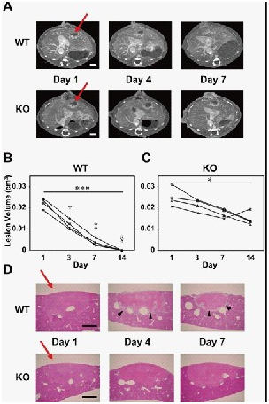
Figure 1. Comparison of liver damage between WT group and plasminogen KO group.
A. CECT scans showed that in WT mice, there was a process of progressive repair of liver injury over time, and this repair was not found in KO mice (arrows indicate areas of liver damage). B. Quantitative analysis of the repair process of liver injury in WT mice. C. KO mice during the two weeks of DAY1 and DAY14 , the change in the volume of the liver injury site. D. H&E staining of the middle part of the liver injury.
Small Animal CT (LCT200): http://?equipid=566719&division=1916
Digital 60m short range laser rangefinder is a laser measurement device which is supposed to replace tape measure, ruler, and other traditional length meauring tool. Laser ruler as its name, it is a electronic ruler which user laser to measure distance.
Our laser measurer has Multiply fuctions:
1. Height measure & Length measure &Distance measure
2. Area Measure
3. Volume Measure
4. Pythagorean Measure
60M Laser Measure,60M Laser Distance Measure,Laser Distance Measure Oem,60M Digitial Laser Distance Measure
Chengdu JRT Meter Technology Co., Ltd , https://www.accuracysensor.com