Literature overview:
Cancer, a malignant tumor, is considered to be incurable. However, if cancer or malignant tumors can be diagnosed early in the disease, they can fight for time and early treatment, thus greatly improving the survival rate of cancer patients. Although immunohistochemistry and other methods have been widely used in the detection of cancer biomarkers, this method has many disadvantages, low sensitivity, and poor judgment in the cancer stage, which can easily lead to misdiagnosis. With the rapid development of nano-biotechnology, new semiconductor nanocrystal quantum dots are gradually applied to the biomedical field. The combination of this technology and tumor biomarkers will study the pathogenesis of cancer, early diagnosis, targeted therapy and prognosis. Have a positive impact.
This paper describes a method for the fusion of single-domain antibodies (sdAbs) based on quantum dots with anti-human epidermal growth factor receptor 2 (HER2) to generate ultra-small luminescent nanoprobes, and successfully applied to the detection of lung cancer and breast cancer. Tumor cells. The researchers first simulated the microenvironment of tumor cell growth in vitro, used the microenvironment to cultivate different lung and breast cancer cell lines, and used Western Blot and ELISA to obtain lung cancer and breast cancer cell lines with different HER2 expression levels. Then, the HER2 single domain antibody was prepared by genetic engineering antibody (see Figure 1): the biomarker HER2 was used to immunize the alpaca, and the lymphocytes in the alpaca serum were used to obtain RNA, and a phage antibody library was constructed, and the activity was high by ELISA. A single domain antibody against HER2, and finally the sequence of the variable region of the single domain antibody is obtained by sequencing. A single domain antibody containing his-tag and cys was constructed by extension PCR (Fig. 2), and engineered sdAbs-SH were obtained by prokaryotic expression. Then by 4-(N-maleimidomethyl)cyclohexane-1-carboxylic acid sulfosuccinimide sodium salt (sulfo-SMCC) or N-(p-maleimido group Phenyl)isocyanate (PMPI) couples sdAbs-SH to quantum dots modified with amino or carboxyl groups (Figure 3). Finally, sdAbs-QDs conjugates were used to detect lung cancer and breast cancer cells with different HER2 expression levels in the microenvironment. The staining of lung cancer and breast cancer was observed by laser confocal microscopy and analyzed by flow cytometry. Figure 5). The article confirmed that compared with the traditional organic dyes Alexa Fluor 488, 568, the staining effect of sdAbs-QDs conjugates was significantly superior in the detection of lung cancer and breast cancer cells with different HER2 expression levels.
Using sdAbs-QDs conjugates, researchers were able to detect HER2 at low levels in cancer cells. This assay combines the excellent fluorescence properties of quantum dots with the extremely high permeability of ultra-small single-domain antibodies, greatly improving detection. Sensitivity. This technology will greatly promote the application of QDs as marker probes in cancer research and diagnosis. The article was published in the journal ACS NANO.
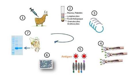
Figure 1: Single domain antibody preparation process diagram
(1) Immunizing alpaca with an antigen of interest.
(2) Peripheral blood mononuclear cells are recovered from blood by density gradient centrifugation.
(3) A gene encoding a single domain antibody was amplified from total RNA reverse transcribed cDNA by RT-PCR and cloned into a phagemid vector.
(4) The foreign gene bank sdAbs were inserted into the coat protein P3 gene to display on the surface of the helper phage.
(5) The sdAb phage display library is adsorbed by the immunizing antigen, the unbound phage is washed away, and the bound phage is eluted for enrichment;
(6) monoclonal screening and identification;
(7) Positive phage sdAbs were infected with non-inhibitory Escherichia coli for soluble secretion expression, purification, and finally subjected to SDS-PAGE analysis.

Figure 2: Single domain antibodies (sdAbs) with his-tag and cys
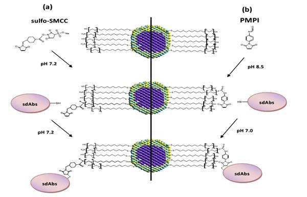
Figure 3: (a) 4-(N-maleimidomethyl)cyclohexane-1-carboxylic acid sulfosuccinimide sodium salt (sulfo-SMCC) and (b) N-(p Schematic diagram of coupling reaction of -maleimidophenyl)isocyanate (PMPI) with quantum dots
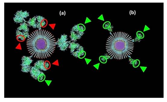
Figure 4: Binding of (a) full length antibody and (b) single domain antibody (sdABs) to quantum dots.
(a) Binding of quantum dots (QDs) to full size antibodies is carried out using carbodiimide chemistry. The binding of the antibody to the quantum dots is randomly oriented, with some antigen binding domains (red arrows) facing the surface of the nanoparticles; only the exposed binding domains are functionally active (green arrows).
(b) Coupling of a Cys residue at the C-terminus of the single domain antibody with a quantum dot ensures that the antigen binding domain of each single domain antibody is exposed, maintaining extremely high functional activity.
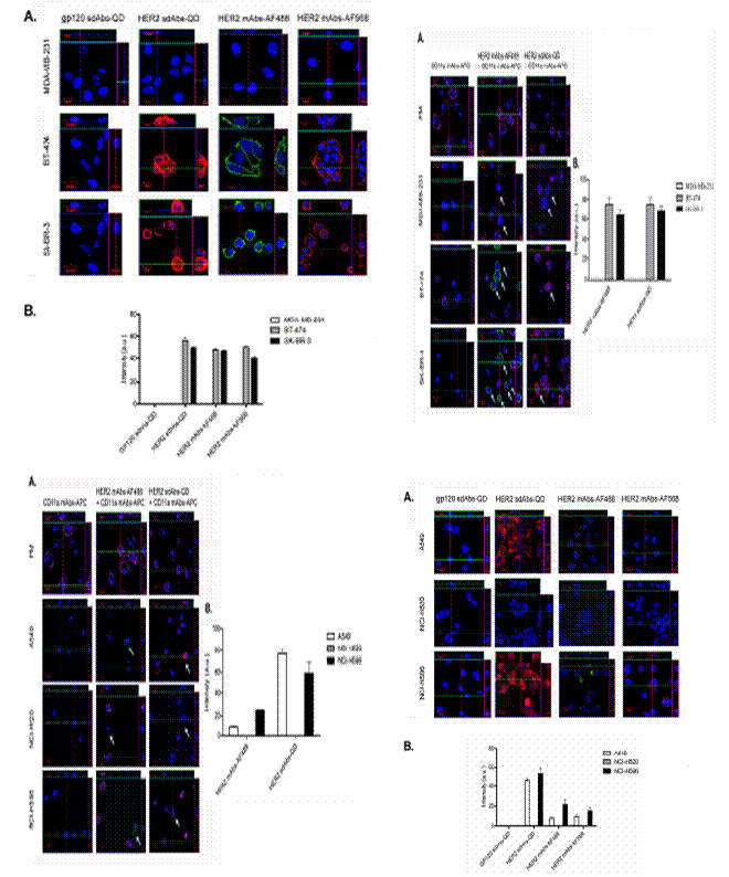
Figure 5: Schematic diagram of detection of lung cancer and breast cancer cells co-cultured with primary macrophages by quantum dots and traditional dye-labeled HER2 conjugates
As customers have higher and higher requirements for animal reproduction and animal disease diagnosis and treatment, traditional diagnostic methods such as visual inspection, stethoscope listening, thermometer inspection, and percussion and hammering can no longer meet the needs of animal husbandry production and veterinary clinics. Today, B-ultrasound is playing a big role in human medical diagnosis, and in the future, B-ultrasound will also play a role in animal medicine. We should promote the use of veterinary ultrasound scanner step by step according to the actual situation, popularize the knowledge of the use of veterinary ultrasound scanner, take this as an opportunity, and use it as a ladder to improve the level of veterinary practice in our country.
Veterinary Ultrasound Scanner,Vet Ultrasound Scanner,Veterinary Ultrasound Machine,Veterinary Ultrasound Equipment
Mianyang United Ultrasound Electronics Co., Ltd , https://www.uniultrasonic.com