Release date: 2016-10-18

The brain can be said to be the most complex organ in the human body, and for scientists, the brain is also one of the most difficult organs to study. The highly complex neural network in the brain makes it impossible to take out tissue for biopsy, so so far, I want to know that the gene expression in the human brain can only be achieved through donation of organs. Recently, researchers at Harvard University in the United States published a newly developed technology in the journal Science Translational Medicine, which can observe the gene activity in the brain in real time. By observing genetic activity in the brain, you can help scientists study how the human brain thinks, feels, and remembers, as well as the signs of diseases such as Alzheimer's and schizophrenia. 
For many neurodegenerative and psychiatric diseases, simple genetic theory does not explain the cause or predict the risk, that is, most patients do not carry disease-causing genes or high-risk factors. This research led to the latest frontier of genetics, epigenetics, which specifically studies whether the same gene is expressed and expressed in different individuals. These gene "switches" in the brain have been found to affect many diseases including Alzheimer's disease, schizophrenia, depression, drug addiction, etc., and even affect the decline of brain function during normal aging. Environmental factors and major events experienced by people, including both physical and psychological trauma, can have a major impact on gene expression. Epigenetics is devoted to studying how genetic "switches" are regulated and how they affect the normal function and disease of organs. Since epigenetics studies the dynamic process of gene regulation, it is impossible to observe these activities in post-mortem tissues, so there is a need for a technique for real-time observation of gene activities.

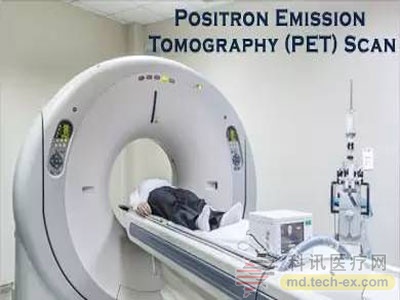
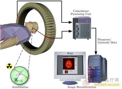
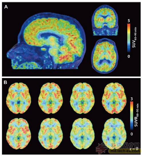
Because of the protection of the brain's hard and opaque skull, it is difficult to study neural epigenetics. There are only a limited number of real-time imaging techniques that can be used to observe brain activity, including positron emission tomography (PET). Traditional brain PET imaging monitors which regions are active by monitoring the distribution of radioactive glucose molecules in the brain. For example, active regions consume more glucose to provide energy. Harvard scientists' newly invented methods for observing brain activity in the brain are also based on PET imaging, which produced a small radioactive molecule called Martinostat. This molecule can cross the blood-brain barrier and specifically bind to brain brain histone deacetylase (HDAC), and the primary function of HDAC is to terminate gene expression. Thus, using Martinitostat-based PET imaging, it is possible to monitor in real time which parts of the brain have stopped expressing.
A team led by Professor Jacob Hooker experimented with eight healthy volunteers at the Massachusetts General Hospital. They proved that the technology was safe and feasible, and found some interesting phenomena. For example, they found that the least active area of ​​gene expression in the brain is the cerebellum and putamen that control motor function, while the most active area is the hippocampus responsible for learning and memory and the amygdala responsible for emotional response. Although it is unclear how to interpret this pattern, one possibility is that the more active the part of the gene is, the more malleable it is, so that these parts can constantly adjust the network between neurons to cope with external changes. Another more important finding is that the distribution of gene expression activity is unexpectedly similar in the brains of these healthy volunteers. The active and inactive areas of a person's brain are almost identical to others. Researchers hope to create a standard "map" of gene activity in the brain, and once a subject's "map" deviates from the standard, it is often a sign of disease. For example, studies have shown that there are a large number of molecules in the hippocampus of Alzheimer's disease that terminate gene expression.

Dr. John Satterlee, head of the Department of Neurogenetics and Epigenetics at the National Institutes of Health, who was not involved in the study, commented: "This is an exciting groundbreaking work that brings us to the unknown of brain gene expression. The field, and depicts the basic blueprint for this new field.†Researcher Professor Hooker said: “This is just the first step in our plan, and we will next try to study the degree of expression of certain genes.†
This real-time imaging technique for observing genetic activity in the brain has broad prospects: one day, this technology will not only predict disease occurrence, but also potentially bring potential treatments. If the molecules that terminate the gene expression are inhibited, it is possible to restore the neuronal activity that is deprived of function in diseases such as Alzheimer's disease and Parkinson's disease, and thus it is expected to restore the normal function of the patient.
Reference materials:
[1] In living color: New technique sees gene activity in human brains
[2] Insights into neuroepigenetics through human histone deacetylase PET imaging
Source: WuXi PharmaTech
Preservation tubes are swabs with disposable virus sampling tubes to collect DNA tests for disposable nasal flocking sterile medical transport.
swabs with disposable virus sampling tubes , to collect DNA tests for disposable nasal flocking sterile medical transport.
Jiangsu iiLO Biotechnology Co., Ltd. , https://www.sjiilogene.com